


Osteoarthritis of the hip joint is a degenerative-dystrophic pathology, which is characterized by destruction of the hyaline cartilage.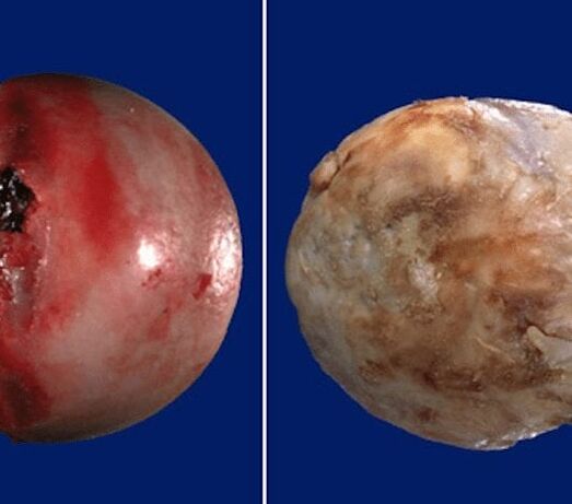 The disease develops gradually, accompanied by pain and decreased movement. In the absence of medical intervention in the initial stage of osteoarthritis, after a few years, atrophy of the femoral muscles occurs.
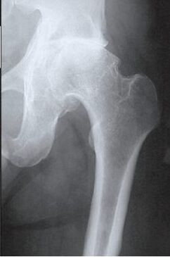 The damaged limb is shortened, and the union of the joint space leads to partial or complete immobilization of the hip joint. Causes of pathology are previous injuries, curvature of the spine, systemic diseases of the musculoskeletal system.
Osteoarthritis is usually detected in middle-aged and elderly patients. Diagnosis is made based on the results of instrumental studies - radiography, MRI, CT, arthroscopy. Treatment of severity pathology 1 and 2 is conservative. If ankylosis is detected or drug therapy is ineffective, surgery (arthrodesis, endoprosthetics) is performed.
Mechanism of pathology development
The hip joint is formed by two bones - the ilium and the femur. The lower part of the ilium is represented by its body, which participates in articulation with the femur, forming the upper part of the acetabulum. During movement, the glenoid fossa is immobile, and the femoral head moves freely. Such a device "hangs" the hip joint allows it to bend, not split, rotate, promotes abduction, traction of the hip. The smooth, supple, elastic hyaline cartilage that wraps around the acetabulum and femoral head ensures unimpeded sliding of the articular structures. Its main functions are redistribution of loads during movement, prevention of rapid consumption of bone tissue.
Under the influence of external or internal factors, cartilage trophism is disturbed. It does not have its own circulatory system - synovial fluid supplies the tissue with nutrients. With osteoarthritis, it thickens, becomes thick.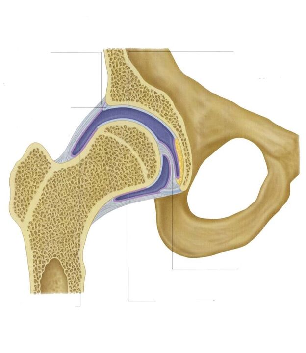 The resulting lack of nutrients provokes drying of the hyaline cartilage surface. It becomes covered with cracks, which leads to permanent tissue microtrauma during flexion or extension of the hip joint. The cartilage becomes thinner and loses its cushioning properties. Bones deform to "adapt" to increasing pressure. And against the background of deteriorating tissue metabolism, destructive and degenerative changes progress.
Causes and provocative factors
Idiopathic or primary osteoarthritis develops for no reason. It is believed that the destruction of cartilage tissue occurs due to the natural aging of the body, the slowing down of recovery processes, a decrease in the production of collagen and other compounds necessary for the complete regeneration of hip joint structures. Secondary osteoarthritis occurs against the background of a pathological condition already present in the body. The most common causes of secondary disease include:
- previous injuries - damage to the ligamento-tendon apparatus, muscle ruptures, their complete separation from the bone base, fractures, displacements;
- joint development disorders, congenital dysplastic disorders;
- autoimmune pathologies - rheumatoid arthritis, reactive, psoriatic, systemic lupus erythematosus;
- non-specific inflammatory diseases such as purulent arthritis;
- specific infections - gonorrhea, syphilis, brucellosis, ureaplasmosis, trichomoniasis, tuberculosis, osteomyelitis, encephalitis;
- dysfunction of the endocrine system;
- degenerative-dystrophic pathologies - osteochondropathy of the femoral head, osteochondritis disecans;
- hypermobility of the joints, due to the production of "super-extending" collagen, provoking their excessive movement, weakness of the ligaments.
Since the cause of the development of osteoarthritis may be hemarthrosis (hemorrhage in the hip joint cavity), provocative factors include disorders of hematopoiesis. Prerequisites for the onset of the disease are excess weight, excessive physical activity, a sedentary lifestyle. Its development is caused by improper organization of sports training, a lack in the diet of foods with a high content of micronutrients, fat-soluble vitamins and water. Postoperative osteoarthritis occurs several years after surgery, especially if it was accompanied by incision of a large amount of tissue. Hyaline cartilage trophism is plagued with frequent hypothermia, living in an unfavorable environment and working with toxic substances.
Osteoarthritis of the hip joint can not be inherited. But in the presence of some innate features (metabolic disorders, skeletal structure), the likelihood of its development increases significantly.
Symptoms
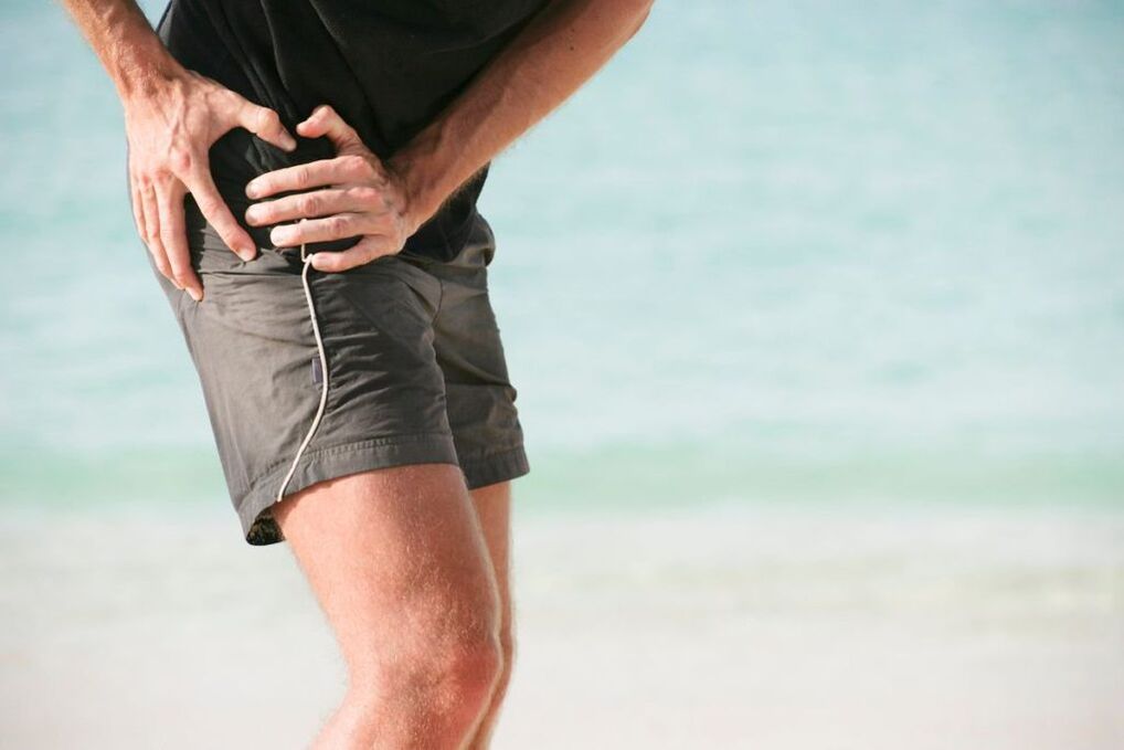
The main symptoms of osteoarthritis of the hip joint are pain when walking in the hip region, radiating to the groin, the knee joint. A person suffers from stiffness of movements, stiffness, especially in the morning. To stabilize the joint, the patient begins to limp, his gait changes. Over time, due to muscle atrophy and joint deformation, the limb shrinks significantly. Another characteristic sign of pathology is limitation of hip abduction. For example, difficulties arise when you try to sit on a bench with your legs apart.
For osteoarthritis of the first severity, periodic pain occurs after intense physical exertion. They are located in the joint area and disappear after a long rest.
With second-degree osteoarthritis of the hip joint, the severity of the pain syndrome increases. Discomfort also occurs at rest, extends to the thighs and groin, increases with weight lifting or increased motor activity. To eliminate pain in the hip joint, a person begins to limp barely visible. Restriction of movement in the joint is observed, especially during abduction and internal rotation of the thigh.
Third-degree osteoarthritis is characterized by persistent severe pain that does not subside day and night. Difficulties arise when moving, therefore, when walking, a person is forced to use a cane or crutches. The hip joint is stiff, has a significant atrophy of the muscles of the buttocks, thighs and legs. Due to the weakness of the abductor femoral muscles, the pelvic bones move to the frontal plane. To compensate for the shortening of the leg, the patient leans from the injured limb when moving. This provokes a strong shift in the center of gravity and an increase in stress on the joints. At this stage of osteoarthritis, pronounced ankle ankylosis develops.
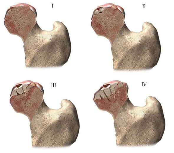
| rank | Radiographic signs |
| First | The changes are not noticeable. The joint gaps are moderate, unevenly narrowed, there is no destruction of the femoral surface. Small bony growths are observed on the outer or inner edge of the acetabulum |
| Second | The height of the joint space is significantly reduced due to its uneven joint. The bony head of the femur shifts upwards, deforms, expands, its contours become uneven. Bone growths form on the surface of the inner and outer edges of the glenoid pit |
| The third | There is a complete or partial union of the common space. The femoral head is strongly enlarged. Numerous bony growths are located on all surfaces of the acetabulum |
Diagnosing
When making a diagnosis, the doctor takes into account the clinical manifestations of the pathology, the anamnesis, the results of an external examination of the patient and instrumental studies. Radiography is the most informative. With its help, the condition of the hip joint, its stage of flow, the degree of cartilage tissue damage is assessed and in some cases the cause of development is determined. If the cervico-diffuse node is enlarged, and the acetabulum is oblique and flattened, then with a high degree of probability it is possible to assume congenital dysplastic changes in articulation. Perthes disease or epiphysis of juveniles is indicated by the disturbed shape of the hip bones. Radiography can detect post-traumatic osteoarthritis, despite the absence of a previous trauma in the anamnesis. Other diagnostic methods are also used:
- CT helps to detect enlarged edges of bone plates, formed osteophytes;
- MRI is performed to assess the condition of connective tissue structures and the degree of their involvement in the pathological process.
If necessary, the inner surface of the joint is examined with arthroscopic instruments. Differential diagnosis is performed to rule out gonarthrosis, lumbosacral or thoracic osteochondrosis. Pain in osteoarthritis can be disguised as clinical manifestations of radicular syndrome caused by nerve blockage or inflammation. It is usually possible to rule out neurogenic pathology with the help of a series of tests. Osteoarthritis of the hip joint is necessarily differentiated from trochanteric bursitis of the hip joint, ankylosing spondylitis, reactive arthritis. To rule out autoimmune pathologies, biochemical studies of blood and synovial fluid are performed.
Medication treatment tactics
Medical treatment aims to improve patient well-being. For this, drugs of different clinical and pharmacological groups are used:
- non-steroidal anti-inflammatory drugs (NSAIDs) - Nimesulide, Ketoprofen, Diclofenac, Ibuprofen, Meloxicam, Indomethacin, Ketorolac. To relieve acute pain, injection solutions are used, and pills, pills, oils, gels help eliminate pain of mild or moderate severity;
- glucocorticosteroids - Triamcinolone, Dexamethasone, Hydrocortisone. They are used in the form of intra-articular blockades in combination with the anesthetics Procaine, Lidocaine;
- muscle relaxants - Baclofen, Tizanidine. They are included in treatment regimens for skeletal muscle spasm, pinchings of sensitive nerve endings;
- drugs that improve blood circulation in the joint - Nicotinic acid, Aminophylline, Pentoxifylline. Patients are prescribed to improve tissue trophism, to prevent disease progression;
- chondroprotectors. Effective only in stages 1 and 2 of osteoarthritis.
Rubbing oils with a warming effect helps eliminate mild pain. The active ingredients of external agents are capsaicin, cinquefoil, camphor, menthol. These substances are characterized by a local irritating, distracting, analgesic effect. Joint compresses with Dimethylsulfoxide, medical bile will help to cope with bloating, morning swelling of the thigh. Patients are recommended a classic massage, acupressure or vacuum for coxarthrosis. Daily exercise therapy is an excellent prevention of further progression of osteoarthritis.
Surgical intervention
With the ineffectiveness of conservative therapy or the diagnosis of a pathology complicated by ankylosis, an operation is performed. It is impossible to restore cartilage tissue to the joint damaged by osteoarthritis without prosthetic surgery, but with the right approach to treatment, adhering to all medical prescriptions, maintaining a correct lifestyle, doing therapeutic exercises, regular massage courses, by taking the right vitamins and nutrition, you can stop the process of damage and destruction of cartilage and hip joints.